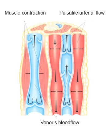
Energy providing substances required by body cells and the oxygen needed to utilize these are sent from the heart to the tissues through arteries. The energy substances and oxygen in this sent blood are used by the tissues, waste materials are added to this blood and they are returned to the heart by the veins in order to be cleaned. As a result of the dilation and convolution of these veins, the illness called “varices” is formed.
 The veins in the body can be examined in two main groups; superficial and deep. Superficial ones are under the skin and are visible to the eye. Deep veins are located between muscles, next to the related arteries and nerves and can’t be seen by naked eye. In order to always have these bloods directed to the heart and not to accumulate them at the legs due to gravity, there are 10-15 valves inside the leg veins. When these valves cannot completely close and blood is leaked down below, blood is accumulated at these veins and their ancillary paths, veins are dilated and the illness what we call varices occur. Although most of these cases are hereditary, it is more common in people who work standing-up (teachers, nurses, doctors, waiters, etc.). It is more frequent in women according to men and varices possibility increases by age.
The disease has stages from showing millimetric capillary vessels to having open wounds in the leg. In early stage patients, there are usually image problems but as the illness progresses, there are complaints such as pain, swelling, sensitivity in the legs. In latter stages leg skin color may darken and have flaking, even non-healing wounds in the legs.
Screening methods are used for diagnosis. Venography or colored Doppler ultrasonography can be used to understand whether there is valve insufficiency at deep, unseen veins. In Venography, a colored substance is given into a vein at ankle level, and films are taken. With today’s modern ultrasonography instruments, use of venography is pretty much reduced. Colored Doppler ultrasonography is a painless, simple method that doesn’t require needles.
Treatment must be performed according to the patient and disease level. In early stage illnesses and for old patients with surgery risks, the progress of the disease can be prevented by merely using varices socks. Below-the-knee varices socks are usually sufficient. At small, 1-3 millimeter varices and capillary vessels, if there are no valve insufficiencies found in Doppler ultrasonography, various treatment methods can be applied. The veins can be entered with special fine needles, and the vein can be closed by giving special substances (sclerotherapy). This method can also be applied by special instruments such as electro-coagulation, radiofrequency and lasers. Several sessions are performed according to the spread of the disease. The patient can walk home after the operation, no rest in bed is necessary. Wearing elastic bandages or varices socks for 1-3 days is sufficient.
In an advanced level disease, methods such as standard surgeries, foam sclerotherapy, radio-frequency and laser ablation can be used. Treatment must be performed according to the patient and disease level. The common goal in each of these methods is to remove the superficial veins with valve insufficiencies or close them to prevent venous load and accumulation. There is no harm for the body in removing or closing these veins, whose valves do not work anyway. Deep veins will do the same job. It is sufficient to have the patient rest at the hospital for one night in each of these cases. Patient can return to normal life in 7-10 days.
A 1-2 centimeter inguinal laceration is performed at a standard surgery. The branches of the superficial vein at the location it is connected to the deep system are tied. With a special cord, the superficial vein is moved to knee level. 5-7 millimeter small lacerations are performed on the expanded varices and these are removed.
Superficial vein is entered by open operation or with Doppler ultrasonography at knee or ankle level in foam sclerotherapy, radio-frequency and laser ablation methods. In foam sclerotherapy, the superficial vein is closed by injecting the medicine, which was mixed and brought to a foam type state. In radio-frequency and laser ablation techniques, the vein is closed by high temperature catheters.


 | +90 212 375 6565 / 6640